
The term Developmental Dysplasia of the Hip (DDH) refers to a group of anomalies of the hip that are present at birth, in which the relationship between the head of the femur and the pelvis (acetabulum) is altered to a greater or lesser extent.
Developmental Dysplasia of the Hip (DDH): what problems does it include?
Children with developmental dysplasia or congenital hip dislocation can present very different pictures:
- from acetabular dysplasia, in which the femoral head and acetabulum have normal relationships but the acetabulum is not shaped sufficiently to adequately contain the femoral head;
- to subluxation, an intermediate condition between dysplasia and dislocation
- to complete dislocation, in which the head of the femur is not correctly centered in the acetabulum and therefore requires rapid treatment to re-establish the correct relationships.
Developmental dysplasia of the hip: what are the causes?
The causes of developmental dysplasia or congenital hip dislocation have not yet been clarified with certainty.
The main role appears to be played by the presence of abnormal postures within the uterus (so-called dislocating postures). In this position, the femoral head is abnormally pressed against the acetabulum, causing abnormal development of the acetabulum itself and laxity of the joint.
For this reason, children with the greatest risk of congenital hip dislocation are those with breech presentation.
Another primary risk factor is a first-degree family history of hip dysplasia, that is, the fact that the mother or father or a brother or sister have suffered from hip dysplasia.
These two factors are the ones that are primarily taken into consideration in those health systems where selective ultrasound screening is performed, that is, reserved only for children with risk factors.
Unfortunately, many studies have shown that hip dysplasia can develop even in the absence of these risk factors.
Other risk factors:
- Female sex. Females are more affected than males. But this doesn’t mean males can’t get hip dysplasia.
- Twinness
- Oligohydramnios: presence of little amniotic fluid;
- Associated malformations (clubfoot, talovalgus foot, myogenic torticollis, etc.). Even for these conditions, the discussion is broad and complex.
- Finally, there may be a genetic predisposition underlying the disease.
Developmental hip dysplasia: manifestations
In the vast majority of cases, developmental hip dysplasia does not present any external manifestations visible to the parents at birth. Only severe cases of complete hip dislocation present with one limb appearing slightly shortened compared to the other and with other obvious signs (e.g., asymmetrical skin folds, associated with the shortening of the limb).
In all other cases, the condition can only be highlighted through a careful clinical evaluation by the neonatologist/pediatrician who examines the baby and through an ultrasound evaluation performed by expert hands.
Developmental dysplasia of the hip: what are the consequences?
If hip dysplasia is not recognized and diagnosed, the evolution can be varied:
- Some very mild cases (mild immaturity, mild dysplasia) may even undergo spontaneous healing in many cases
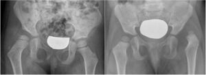
- True hip dysplasia, however, can progressively worsen. In more severe cases, this can lead to progressive hip dislocation, which can occur more or less rapidly. This can manifest itself, for example, when a child begins to walk, with a noticeable limp and shortening of the limb. In less severe cases, however, progressive dislocation does not develop; the femoral head remains in place, but due to a lack of perfect congruence between the femoral head and the acetabulum, the joint functions poorly and wears out more rapidly, thus developing premature osteoarthritis (which often leads to the need for a hip replacement at a young age).
- Hip dislocation if left untreated generally leads to shortening of the limb and significant lameness.
Developmental dysplasia of the hip: diagnosis
The two fundamental elements for the diagnosis of hip dysplasia are:
- Clinical examination by a neonatal specialist/pediatrician capable of adequately performing the maneuvers
- Hip ultrasound performed by expert hands.
1) During the examination, the examiner must be able to recognize the various typical signs of hip dysplasia: instability, reduced abduction, shortening, asymmetrical skin folds, etc.
Certain maneuvers are essential for recognizing the most obvious signs of hip dysplasia/dislocation: the Ortolani maneuver and the Barlow maneuver. It is essential that these maneuvers be performed at birth and during initial health checks, and recorded in the dog’s medical record. These maneuvers are not easy and must be performed by experienced personnel.
Unfortunately, not all cases of hip dysplasia are positive on clinical examination. For this reason, it is essential to enhance the diagnosis with an additional, non-invasive tool: ultrasound, performed by experienced personnel.
2) Hip ultrasound can accurately diagnose the morphology of the hip joint and recognize all degrees of the disease, from the mildest cases that may escape clinical examination to the most severe. For this reason, it is the cornerstone of hip dysplasia diagnosis.
Key concept : simply performing a hip ultrasound isn’t enough to give you peace of mind. It’s essential that the ultrasound be performed by experienced personnel and following precise criteria, as described by its creator, Graf.
Hip dysplasia: when should an ultrasound be performed?
The current recommendations of the Italian Society of Pediatrics and Pediatric Orthopedics recommend performing an ultrasound on all newborns between 6 and 8 weeks of age.
Developmental dysplasia of the hip: treatment
Hip dysplasia (or congenital hip dislocation) comprises a set of developmental abnormalities of the hip that lead to a change in the morphology and relationship between the femoral head and the pelvis. Treatment requires centers with specific experience.
Our center has an outpatient clinic that handles all aspects, from ultrasound diagnosis to the application of braces or casts, to surgical procedures (closed/open reduction, femoral and pelvic osteotomies, etc.)
Please refer to other sheets for specific aspects: