
Proximal Femoral Focal Deficiency


In Congenital Femoral Deficiency (CFD), it is important to evaluate the condition of the hip, which may appear completely normal or exhibit mild to severe pathological features. These alterations are generally referred to as Proximal Femoral Focal Deficiency (PFFD) and use the Aitken classification. Treatment of Proximal Femoral Focal Deficiency requires centers with experience.
We use modern and excellent surgical techniques to intervene on the pathology “Proximal Femoral Focal Deficiency

In Congenital Femoral Deficiency (CFD), it is important to evaluate the condition of the hip, which may appear completely normal or exhibit mild to severe pathological features. These alterations are generally referred to as Proximal Femoral Focal Deficiency (PFFD) and use the Aitken classification.

The pelvis may present dysplasia : the inclination of the acetabular roof (where the head of the femur rests when the patient is standing) is insufficient, posing a risk of the femur gradually being pushed towards dislocation.
The proximal femur may also present variable conditions: in mild cases, it presents a varus alteration of its normal angle (between the diaphysis and the neck), called coxa vara. The condition can be more or less severe depending on the severity of this angle (greater or less than 90°).
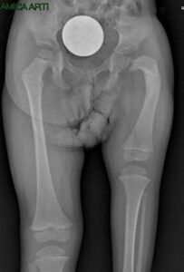
Congenital Femoral Deficiency with coxa vara (with an angle of approximately 90°)

In intermediate cases, it presents a pseudoarthrosis : the segment of the head and that of the diaphysis are not continuous with each other, in between there is a pathological, non-ossified tissue, which does not provide stability to the segment.

Proximal femoral focal deficiency type 2, Paley, Aitken B (followed elsewhere). a) X-ray at 5 months of age. No reconstructive procedure performed; b, c) CT with 3D reconstruction at 7 years of age.

In severe cases of Proximal Femoral Focal Deficiency , there is severe subversion with the absence of the entire proximal femur and part of the diaphysis.
In these cases, an MRI scan is useful to better understand the localized condition. In some cases, for example, a pseudarthrosis is mistakenly diagnosed in conditions of simple delayed ossification, which improves spontaneously over time.

Proximal femoral focal deficiency with severe shortening of the femur. The proximal portion of the femur, the femoral neck and head, are not ossified, so it is not possible to fully understand the pathological condition and establish a plan with a simple x-ray.

See the fact sheet “Congenital Femoral Deficiency”.

It is important to contact centers with specific experience, such as OrthoChildren Center.

Techniques for pathology Proximal Femoral Focal Deficiency

SUPERKnee procedure: Knee reconstruction for congenital hypoplasia

SUPERHip procedure: hip reconstruction for congenital hypoplasia