
What is an exostosis?
Exostosis (or osteochondroma) is a very common benign lesion in children, characterized by the formation of a hard protuberance that arises from the bone surface, usually near one end of a bone segment. More precisely, exostosis is a hamartoma of the growth plate, that is, a portion of cartilage that normally contributes to bone growth. In this case, it grows in an abnormal location and direction . However, it behaves similarly to the rest of the cartilage: starting from the cartilage at the apex of the protuberance (cartilaginous cap), it tends to progressively become bone (enchondral ossification), thus causing the exostosis to grow .

Solitary exostoses are most commonly located on long bones (distal femur, proximal humerus, proximal tibia), at the metaphysis, that is, immediately above the growth plate of that bone segment. They can vary greatly in shape and size; the base can be broad (sessile) or narrow (pedunculated).
They tend to grow at a variable rate as the patient grows. Once the patient has reached the end of growth, the exostoses stop growing.
Can solitary exostoses transform into a malignant lesion?
The risk of transformation into a malignant lesion (peripheral chondrosarcoma) is extremely low (below 1%) and in any case does not occur in pediatric age.
Exostosis: what are the symptoms?
Typically, the only symptom of exostosis is the sensation of a mass , which the patient often notices by chance. This mass feels hard, extends from the bone surface, and is painless.
In some cases, as the exostosis grows, it compresses surrounding structures (vessels, nerves, etc.), causing discomfort.
Hereditary multiple exostoses (HME)
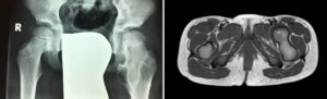
Hereditary multiple exostoses (HME)) is a rare condition (incidence of 1-2 cases per 100,000 inhabitants) characterized by the formation of exostoses in different parts of the body.
These lesions are mostly located in the metaphyses of long bones, but frequently also in the scapula, ribs, and other areas.

From a genetic point of view, exostotic disease is characterized by autosomal dominant genetic transmission (see other discussions for a more detailed analysis of the genetic aspects).
Depending on the anatomical location , the radiographic morphology of exostoses tends to be different: for example, at the level of the lower (distal) part of the femur, exostoses tend to take on a pedunculated appearance in the proximal direction, at the proximal tibia they tend to take on a pedunculated appearance in the distally direction, at the level of the fibula and humerus they tend to take on a sessile appearance, at the level of the ulna they tend to locate in the distal part of the segment (towards the wrist) and cause shortening of the bone.
The clinical picture of multiple exostoses disease is characterized by:
- multiple swellings starting from the bone plane;
- functional limitation (variable from case to case);
- aesthetic damage (variable from case to case);
- short stature ;
- limb length asymmetry ;
- angular deformities of the limbs
- possible malignant transformation .

The clinical presentation phenotype of multiple exostosis disease can be highly variable , from cases with numerous exostoses and significant clinical presentation to very mild cases.
Hereditary multiple exostoses (HME): follow-up of the disease
The continuous and simultaneous growth of lesions throughout the growth period must be considered , so it is important not to overdo radiographic examinations (to limit radiation exposure) and to avoid surgically treating all lesions.
It is advisable to undergo clinical check-ups every 6–8 months during growth. Periodic clinical photographs may be helpful to monitor the progression of the condition. Radiographic examinations may be useful in the event of significant changes in the condition (obvious increase in volume, onset of symptoms, etc.). MRI and CT scans may be performed if there is any suspicion of transformation or to evaluate possible compression of an exostosis on nearby vascular-nervous structures.
Risk of malignant transformation :
The incidence of malignant transformation varies greatly depending on the case study (0.5-25%) and occurs more easily in adulthood , affecting the limbs, trunk, and around the knee. It generally manifests itself with an increase in the size of a mass that appears painful .
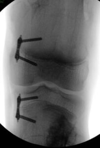
Surgical treatment of exostoses
As mentioned, indications for surgery must follow very specific principles. The main indications for exostosis removal are:
- functional limitation
- pain (rare)
- compression on nearby vessels and nerves
doubtful malignant transformation of an exostosis
There are also other surgical procedures to consider for the possible clinical pictures that may manifest in multiple exostotic disease:
Exostosis: surgery to correct deformities
The growth of exostoses can cause limb deformation through various mechanisms (compression, growth suppression, etc.) .
These deformities can be corrected with various techniques:
- osteotomies
- hemi-epiphysiodesis
- lengthening with axis correction
Please refer to the respective factsheets for explanations of the surgical techniques.
Exostoses: surgery to correct heterometry
By the same mechanisms, patients with multiple exostotic disease may present length differences (LLD) of the upper and lower limbs or relative length differences between the forearm bones.
There are several surgical options to consider:
- hemi-epiphysiodesis to slow the growth of the longer limb
- lengthening of shortened bone segments
Please refer to the data sheets relating to the individual techniques.

OrthoChildren Center performs all these procedures