Sheet Equinus foot treatment
Important:
- distinguish the different types of equinus foot (flexible, rigid, initial contact, swing, masked, equinovarus, equinovalgus, etc): see fact sheet
- Include equinus foot assessment within a comprehensive evaluation of functional surgery in patients with cerebral palsy (see fact sheet).
- use the principles of multilevel surgery in cerebral palsy (see fact sheet)
- know advantages and risks of multiple fibrotomy and SPML: see fact sheet
Equinus foot and surgical decision: examples and risks
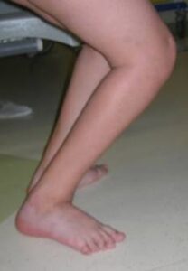
- For example, there are cases in which the foot first contacts the ground with the toe, but only because the knee is flexed (so-called apparent equinus foot). In this case, surgery performed on the foot will only worsen the situation, while proper surgery should address the knee deformity.
- In other cases, equinus foot is rigid, but is associated with marked weakness of the extensor muscles of the foot and toes (which normally keep the foot raised while the limb is moving forward and the contralateral limb is on the ground). In these cases, corrective surgery for equinus foot will correct the stiffness of the foot but will have no effect on the weakness of the extensors. Therefore, post-operatively, the tendency to have the toe pointing downward (swing equinus foot) during the swing phase of the step will persist. This aspect must be adequately discussed with the parents before surgery, and if necessary, appropriate therapeutic measures must be planned (physiotherapy, braces, tibialis anterior tensioning, tendon transfers, etc.).
- Other children have a rigid equinus foot , but also show in their natural history a marked tendency to develop triceps insufficiency, that is, a progressive weakness of the muscle, which puts them at risk of developing progressive knee flexion deformities (crouch gait, see fact sheet) and progressively losing the ability to walk. An inexperienced surgeon will try to correct this equinus as much as possible, when instead he should be aware of such situations and try to take measures to limit (as much as possible) the possible negative consequences.
Surgical treatment: what interventions?
Treatment options for equinus foot include conservative treatments (physiotherapy, bracing, insoles, botulinum toxin injections, etc.) and surgical treatments.
While a description of the former is beyond the scope of this article, we will focus on the possible surgical interventions that can be offered to families for the treatment of equinus foot.
All possible surgical techniques for correcting equinus foot are primarily aimed at lengthening one structure: the triceps surae muscle (or rather, the muscle-tendon unit) . This muscle is formed by the union of three heads: the two gastrocnemius muscles (which insert proximally above the knee, on the femoral condyles: a biarticular muscle) and the soleus muscle (which inserts proximally below the knee: a monoarticular muscle). These muscles join together at the bottom to form the Achilles tendon, which inserts onto the posterior aspect of the heel. The gastrocnemius muscles are more superficial than the soleus muscle (and characterize the profile of the calf).
Shortening or contracture of the triceps surae causes equinus and may be an indication for surgery to lengthen this structure. This contracture can involve the gastrocnemi and soleus muscles equally, but it can also affect these muscles differently (mostly greater contracture of the gastrocnemi, lesser contracture of the soleus).
It is therefore essential for the surgeon to distinguish which part has the contracture and perform surgical procedures targeted to the structures actually involved, avoiding excessively lengthening (and weakening) structures that are not contracted.
To this end, the Silverskiold test is used , which consists of measuring the degree of equinus with the knee initially flexed and subsequently with the knee extended. This test can also be reassessed by the surgeon in the operating room, under general anesthesia.
Obviously, the segmental examination must also include other assessments (evaluation of spasticity, selective motor control, etc.).
Once the necessary assessments have been made, the surgeon will be able to plan the type of surgery for equinus foot . The techniques are divided into surgical levels, that is, based on the anatomical area of the triceps surae they address. Therefore, considering the most commonly used techniques, the following levels can be distinguished:
- a proximal area (in the upper part of the leg), where the muscular belly of the gastrocnemi is present; deeper down is the muscular belly of the soleus muscle (covered by its aponeurosis);
- a little further distally, the muscle bellies of the gemelli end in a tendon (the so-called distal gastrocnemius tendon); deeper still is the muscle belly of the soleus muscle (covered by its aponeurosis);
- the aponeurosis of the soleus and the distal tendon of the gastrocnemius join to form the so-called cojoint gastrocnemius-soleus tendon; the most distal part of the soleus muscle is still present at depth;
- finally at the bottom the muscle fibres end and only the tendinous structure of the Achilles tendon remains.
Based on the above levels, the following surgical procedures are therefore distinguished:
- The Baumann technique: involves lengthening the aponeurosis covering the gemelli muscles (and possibly the soleus) in the proximal part of the leg; the muscle fibers of the gastrocnemius and soleus are left intact;
- The Strayer technique involves lengthening (or simple tenotomy) of the distal gastrocnemius tendon, proximal to the point where it joins the soleus aponeurosis; it is possible to associate lengthening of the soleus aponeurosis at the same level (deeper);
- The Vulpius and Baker techniques involve lengthening the gastrocnemius-soleus cojoint tendon, keeping the soleus muscle fibres intact in depth;
These techniques are often generically grouped together as muscle recessions or myotendinous lengthenings, having in common the characteristic of allowing the lengthening of the muscle-tendon unit without interrupting its continuity. The continuity of the muscle fibers is maintained intact, but they stretch following the procedure. This would ensure less post-operative muscle strength loss, in addition to the targeted action on the muscle actually involved;
- Achilles tendon lengthening techniques: These are particularly indicated when the equinus deformity is severe and the shortening involves both the gastrocnemius and soleus. This procedure can be performed:
- open, that is, by making a skin incision, visualizing the tendon and lengthening it as necessary by performing a Z on the tendon ( Z-shaped lengthening )
- Percutaneous technique : by means of multiple millimetric cuts on various sides of the tendon, the latter is weakened until it yields and stretches. Generally, two (White technique) or three cuts (Hoke technique) are made.
Postoperatively, a cast (a cast from below the knee to the toes) is generally applied, which will be worn for 3 to 6 weeks, depending on the technique used and associated procedures.
Any changes or the need for postoperative splinting will be discussed with the parents when planning the procedure.
OrthoChildren Center has experience with all these procedures.
Patients from abroad: can OrthoChildren Center treat foreign patients?
Yes, many patients come from all countries (Europe and USA):
- the surgical equipe has a wide experience with this condition
- an intensive rehabilitation program can be included
- the procedure is less expensive than in USA and other countries
- Families take advantage of this opportunity to combine a trip to the beauties of Italy