Sheet Equinus foot and ankle
Important:
- Include equinus foot assessment within a comprehensive evaluation of functional surgery in patients with cerebral palsy (see fact sheet).
- use the principles of multilevel surgery in Cerebral Palsy (see fact sheet)
Equinus foot in cerebral palsy: definition and types
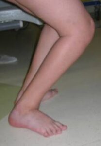
Equinus foot is one of the most common deformities in children with cerebral palsy, as well as one of the most complex.
There are numerous definitions of equinus foot : to simplify, we can define equinus as a foot whose longitudinal axis forms an angle greater than 90° with the longitudinal axis of the leg (in the position of maximum correction); in other words, the toe remains pointed downward and dorsiflexion (upward) of the ankle is impossible. This results in an alterated support (which will occur with the toe on the ground) and gait pattern.
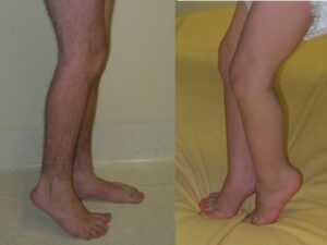
This alteration may be present in only one foot (unilateral equinus foot , as generally occurs in forms of hemiparesis) or in both (bilateral equinus foot, with symmetrical or asymmetrical involvement).
Equinus, equinovarus, and equinovalgus foot
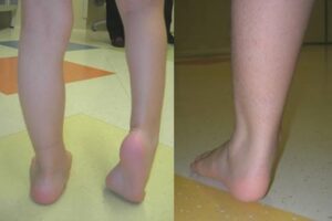
In pure equinus forms , the axis of the hindfoot is aligned with the axis of the leg: in other words, looking from behind the patient, the heel is on the same axis as the leg and shows no obvious deviations inward or outward.
If such deviations are present, the foot will have to be defined differently (equinovarus foot or equinovalgus foot) and other aspects will have to be taken into consideration in the evaluation and treatment (see other factsheets).
Equinus foot: classification
The characteristics of equinus must be carefully evaluated and described.
Therefore, a distinction is made between:
- a rigid equinus foot (in which the deformity is rigid and uncorrectable) and
- a dynamic equinus foot, in which the deformity manifests itself during walking (as a consequence of neurological and muscular mechanisms), but is completely correctable in the evaluation carried out with the patient on the table.
Depending on the phase of the gait in which this equinus occurs, we distinguish:
- Initial contact equinus, midstance equinus, push-off equinus, swing equinus, etc., and each typology will suggest certain problems and correspond to certain treatments.
Further distinctions can also be made: functional, masked equinus, etc.
Equinus foot and surgical decision: examples and risks
- For example, there are cases in which the foot first contacts the ground with the toe, but only because the knee is flexed (so-called apparent equinus foot). In this case, surgery performed on the foot will only worsen the situation, while proper surgery should address the knee deformity.
- In other cases, equinus foot is rigid, but is associated with marked weakness of the extensor muscles of the foot and toes (which normally keep the foot raised while the limb is moving forward and the contralateral limb is on the ground). In these cases, corrective surgery for equinus foot will correct the stiffness of the foot but will have no effect on the weakness of the extensors. Therefore, post-operatively, the tendency to have the toe pointing downward (swing equinus foot) during the swing phase of the step will persist. This aspect must be adequately discussed with the parents before surgery, and if necessary, appropriate therapeutic measures must be planned (physiotherapy, braces, tibialis anterior tensioning, tendon transfers, etc.).
- Other children have a rigid equinus foot , but also show in their natural history a marked tendency to develop triceps insufficiency, that is, a progressive weakness of the muscle, which puts them at risk of developing progressive knee flexion deformities (crouch gait, see fact sheet) and progressively losing the ability to walk. An inexperienced surgeon will try to correct this equinus as much as possible, when instead he should be aware of such situations and try to take measures to limit (as much as possible) the possible negative consequences.