
Fibular hemimelia in children
Fibular hemimelia is a birth defect that belongs to the category of deficiency of the leg (i.e., the segment from the knee to the ankle), affecting the development of the bones that constitute it: the tibia (the main, weight-bearing bone) and the fibula (the lateral, thinner bone).
They are also called longitudinal hemimelias and are divided into:
- those that mainly affect the tibia: Tibial Hemimelia
- those that mainly affect the fibula: Fibular Hemimelia.
Fibular Hemimelia: what is it
In fibular hemimelia, the fibula is hypoplastic (short, poorly developed) or completely absent. This is associated with varying degrees of alterations of the tibia, knee, ankle, and foot, and a shortening of the leg segment.
The severity of the clinical picture can vary greatly, from very mild to very severe cases. It is often associated with involvement of the femur ( Congenital Short Femur, see related factsheet ).

This is a non-hereditary condition , the cause of which is unknown. The only exception is rare cases of bilateral fibular hemimelia, which may be inherited autosomal dominantly.
Fibular hemimelia: clinical problems
This is a complex condition. Treatment must consider all aspects of the disease. The main issues to consider are:
Leg shortening (short leg)
In patients with fibular hemimelia, one leg is already shorter than the other at birth, to a greater or lesser extent. However, the fundamental characteristic that must be clearly explained to the family is that this difference in length will not remain constant, but will tend to progressively increase throughout growth.
In fact, what remains constant , from birth to the end of growth, is the percentage of shortening compared to the healthy leg. For example, if at birth, compared to the healthy leg that is 12 cm long, the leg affected by fibular hemimelia is 1.5 cm shorter (therefore 10.5 cm), the percentage of shortening will be 12.5%. Over time, the shortening will not remain stable at 1.5 cm, but the percentage will remain stable, 12.5%. If we assume that at the end of growth the healthy leg can become on average 40 cm long, we can calculate a hypothesis of shortening of the leg, at the end of growth, of 5 cm (=12.5% of 40 cm).
This approach allows us to establish a treatment plan for patients with fibular hemimelia from the initial assessments, allowing for recovery of the shortening at the end of growth.
However, it is important to consider, especially in assessments performed on very young children, the possibility of calculation errors, primarily related to errors in calculations and measurement of x-rays. Therefore, in subsequent visits, it will be possible to improve the accuracy of these measurements and the corresponding calculations.
There are several ways to perform these calculations. Recently, applications based on Professor Dror Paley’s Multiplier method have also been developed to facilitate the calculations.
In fibular hemimelia, the expected shortening at the end of growth (and the associated shortening at birth) varies greatly, from mild cases with 2-3 cm of shortening to severe cases with over 20-25 cm of shortening , to which associated shortening of the femur may be added (see the relevant factsheet).
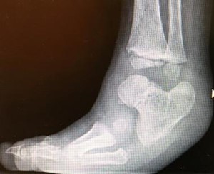
Ankle deformity
In fibular hemimelia, the ankle can be affected in a very variable manner, from no changes to mild valgus to severe articular alteration. Consequently, the joint can present as stable or unstable and deformed in equinus, varus, or valgus (see Paley classification below).
Part of the problem stems from the lack of the normal stabilizing function of the distal part of the fibula (lateral malleolus). Without this, the ankle will tend to show instability or outward subluxation.
Part of the problem, however, stems from the articulation between the talus and the calcaneus (subtalar), which is often malformed or fused , and this fusion may be present in an altered direction.
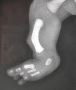
Foot deformity
In fibular hemimelia, the foot can be affected to varying degrees. The lateral aspect of the foot is usually primarily affected, precisely because the initial pathological process (during pregnancy) affects the lateral development of the leg (fibula) and foot.
This frequently results in the absence of the toes and lateral metatarsals, to varying degrees (four-toed foot, three-toed foot, etc.).
This is associated with numerous other possible malformations, which can significantly affect the position of the foot.

Tibial deformity
Fibular hypoplasia is often associated with a deformity of the tibia at the diaphysis level, with a tendency toward valgus and procurvation.
A typical skin umbilication is often present at the apex of the deformity , but this has no pathological significance.
Knee instability
Many patients with fibular hypoplasia present with hypoplasia or complete absence of the cruciate ligaments (anterior and/or posterior).
This is an aspect that is often overlooked without sufficient experience with this condition, but it is crucial in the treatment process.
This instability generally does not cause any problems in young children, but it can in adolescence and adulthood. Above all, it should be considered in the case of lengthening procedures.
Genu valgum
In fibular hemimelia, knee valgus deviation is very common. Poor development of the lateral femoral condyle or the lateral aspect of the proximal tibial epiphysis may be the cause.
Furthermore, following lengthening procedures, valgus deviation may increase. Therapeutic procedures may be necessary for this condition.

Classification of fibular hemimelia
There are numerous classifications. The most widely used classification is that of Achterman in Kalamchi:
- Type 1A: fibula present, its proximal epiphysis is distal to the proximal physis of the tibia
- Type 1B: Fibula partially absent, 30 to 50% of its length; distally the fibula is present but does not support the ankle
- Type 2: Complete absence of the fibula
Among the various classifications, also due to our connection with the School of Pediatric Orthopedics of the Rizzoli Orthopedic Institute, we would like to recall the classification of Dal Monte and Donzelli (O. Donzelli, L. Valdiserri: classification and long-term prediction of lower limb length discrepancies: “Current news in orthopedic surgery” 1982: 573-579):
- Type 1: Miniature fibula: slightly valgus-pronated foot, maximum percentage deficit 15%
- Type 2: hypoplastic-aplastic fibula: tibial procurvation, absence of external rays of the foot, mild femoral hypoplasia, maximum percentage deficit 35%
- Type 3: aplastic fibula, hypoplastic femur: as above but with significant femoral hypoplasia, maximum percentage deficit 60%.

More recently, a new classification has been introduced, that of Professor Dror Paley (J Child Orthop 2016: Surgical reconstruction for fibular hemimelia), which instead primarily considers problems and deformities of the ankle and subtalar joint. This distinction is very useful for establishing a therapeutic reconstruction plan.
- Type 1: Stable ankle. The ankle is often normal, the fibula is shorter at its proximal part.
- Type 2: Dynamic valgus. The ankle goes into valgus, often due to a raised lateral malleolus. Equinus is not present.
- Type 3: Fixed equinus and valgus deformity . The deformity is caused by problems at the ankle (3A), subtalar joint (3B), or both (3C).
- Type 4: Fixed equinus and varus deformity. There is a fusion of the talus and calcaneus, causing the foot to be in varus. This condition can be mistaken for congenital clubfoot.
